
Imaging studies – what are they and what role do they play in modern diagnostics?

 4 lipiec 2026
 4 lipiec 2026
- What are imaging studies and why are they so important?
- What is ultrasonography (USG)?
- What is the significance of radiography (X-ray)?
- When is computed tomography (CT) used?
- How does magnetic resonance imaging (MRI) work?
- What role do imaging studies play in monitoring treatment?
- Are all imaging studies safe?
- Questions and Answers Section
Do you need a prescription, sick leave, or medical consultation?
Order nowImaging studies are a fundamental tool of modern medicine, allowing for non-invasive assessment of internal organs, bones, and soft tissues. They enable early detection of diseases, monitoring of treatment, and planning of therapy based on objective anatomical and functional data.
- Imaging studies are the cornerstone of medical diagnostics.
- Different imaging methods are based on different physical phenomena.
- The choice of examination depends on the type of suspected pathology.
- Scientific studies confirm their crucial role in monitoring treatment.
See also: Magnetic Resonance Imaging (MRI) – learn the most important aspects of this examination.
What are imaging studies and why are they so important?
Imaging studies are a group of diagnostic procedures that allow for obtaining images of the anatomical structures of the body without the need for surgical intervention. They are one of the foundations of medical diagnostics in all clinical fields.
Their significance arises from the ability to detect pathological changes early, often before clinical symptoms appear. Population and clinical studies show that the use of imaging significantly improves diagnostic accuracy and shortens the time to implement appropriate treatment.
Medical imaging is also essential in assessing the effectiveness of therapy and planning further management.
What is ultrasonography (USG)?
Ultrasonography is an examination that uses ultrasound to image internal structures of the body. High-frequency sound waves bounce off tissues, creating real-time images.
USG has wide applications in assessing abdominal organs, the thyroid, blood vessels, and in obstetric diagnostics. Scientific studies confirm its high safety, as it does not involve exposure to ionizing radiation.
The advantages of ultrasonography include availability, the possibility of repeated examinations, and dynamic assessment of the structures being examined.

What is the significance of radiography (X-ray)?
Radiography is one of the oldest and still commonly used imaging methods. It is based on the use of X-rays, which penetrate tissues to varying degrees.
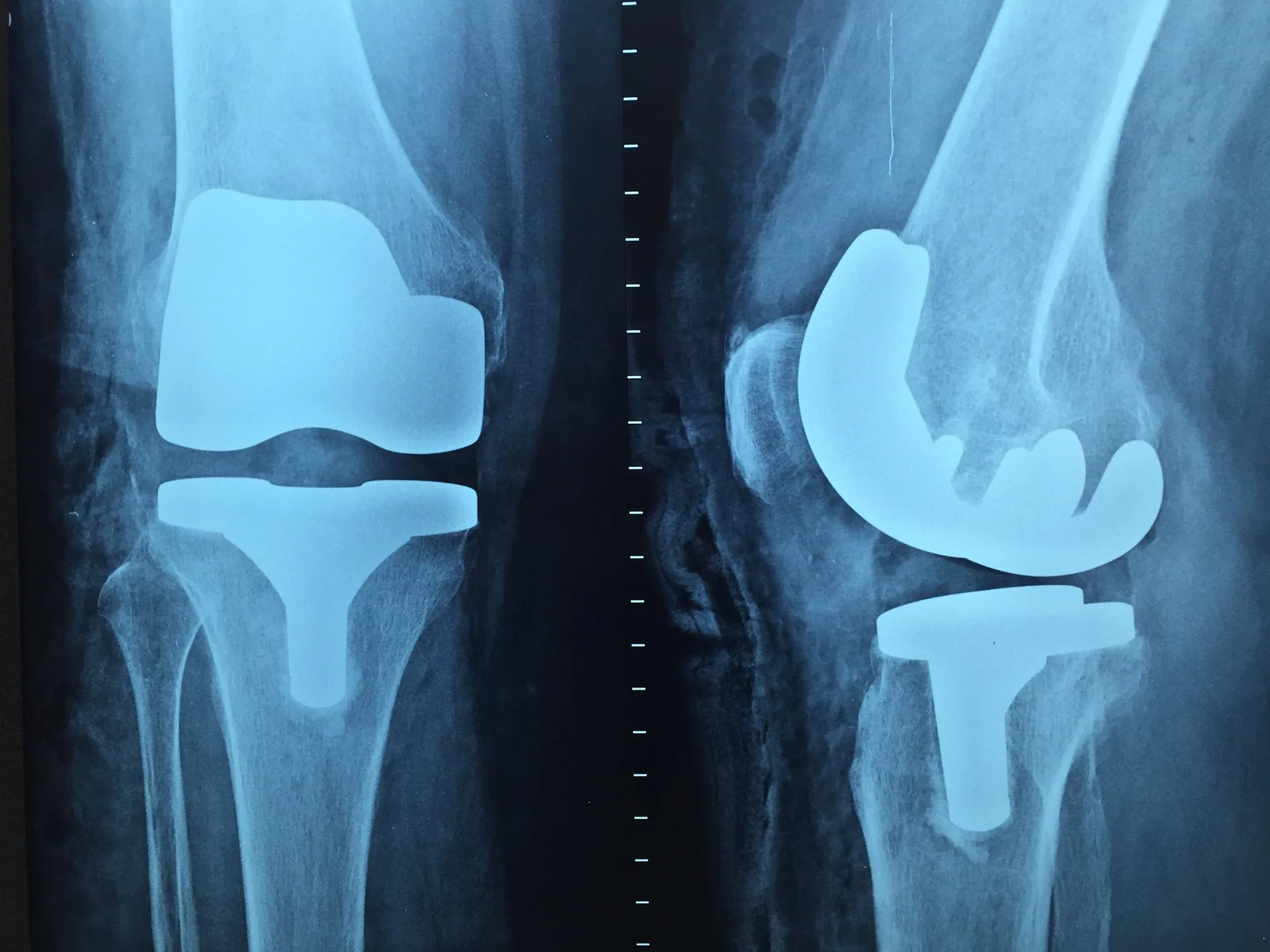
X-ray is particularly useful in diagnosing the skeletal system, allowing for the assessment of fractures, degenerative changes, and postural defects. Studies confirm its high diagnostic value with relatively low radiation exposure when performed according to radiological protection principles.
This method is also used in assessing the chest, especially in lung diseases.
When is computed tomography (CT) used?
Computed tomography is an advanced imaging technique that uses X-rays to create cross-sectional images of the body. It allows for very accurate assessment of internal organs and anatomical structures.
Scientific studies indicate that CT is particularly useful in diagnosing injuries, tumors, and vascular diseases. Due to its high resolution, it enables the detection of even small pathological changes.
Due to the radiation dose, this examination is performed only with clear clinical indications.
How does magnetic resonance imaging (MRI) work?
Magnetic resonance imaging uses a magnetic field and radio waves to obtain detailed images of soft tissues. This method does not use ionizing radiation.
MRI is the gold standard in diagnosing the nervous system, joints, and soft tissues. Studies confirm its high sensitivity in detecting inflammatory, neoplastic, and degenerative changes.
Due to the strong magnetic field, the examination requires appropriate patient qualification, especially in the presence of implants.
What role do imaging studies play in monitoring treatment?
Imaging studies are essential in assessing the effectiveness of ongoing therapy. They allow for an objective comparison of the state before and after treatment.
In oncology, they enable the assessment of tumor response to therapy, and in orthopedics, they monitor the bone healing process. Clinical studies show that regular imaging monitoring improves treatment outcomes and reduces the risk of complications.
Imaging also forms the basis for decisions regarding the continuation or modification of therapy.

Are all imaging studies safe?
The safety of imaging studies depends on the method used. Ultrasonography and magnetic resonance imaging are considered very safe, while methods using X-ray radiation require rational use.
Epidemiological studies emphasize that the risk associated with radiation is small compared to the diagnostic benefits when examinations are performed according to recommendations.
Proper patient qualification and the selection of the appropriate method are crucial.
Questions and Answers Section
Are imaging studies always necessary for diagnosis?
Not always, but in many cases, they significantly increase the accuracy of diagnosis.
Is magnetic resonance imaging safer than computed tomography?
Yes, it is.because it does not use ionizing radiation.
Is one imaging study enough to assess treatment?
Follow-up studies are often needed at specific intervals.
Sources
- Brenner DJ, Hall EJ. Computed tomography — an increasing source of radiation exposure. New England Journal of Medicine.
- Hricak H, et al. Role of imaging in patient management. Radiology.
- Smith-Bindman R, et al. Ultrasonography versus CT in diagnostic imaging. JAMA.
