
Magnetic Resonance Imaging (MRI) – Discover the Key Aspects of This Examination

 4 lipiec 2026
 4 lipiec 2026
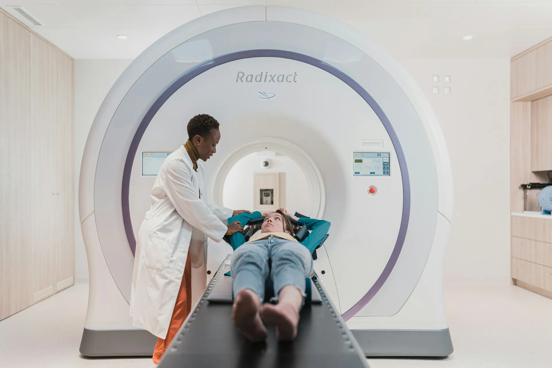
- What is magnetic resonance imaging (MRI) as an imaging test?
- How does magnetic resonance imaging work and what is the role of the magnetic field?
- What structures are best evaluated by magnetic resonance imaging (MRI)?
- When does a doctor refer a patient for magnetic resonance imaging?
- Does magnetic resonance imaging always require contrast?
- What are the limitations and contraindications for MRI?
- Q&A – frequently asked questions by patients about MRI
Do you need a prescription, sick leave, or medical consultation?
Order nowMagnetic resonance imaging (MRI) is one of the most accurate imaging tests used in modern medicine. In medical diagnostics, it allows for a detailed assessment of the internal structures of the body without the use of ionizing radiation, making it a key tool in diagnosing many diseases.
- Allows for precise evaluation of soft tissues
- Is a fundamental test in neurology and orthopedics
- Does not use X-ray radiation
- Is used according to clearly defined medical indications
See also: When is a CT scan needed and what is it actually for?
What is magnetic resonance imaging (MRI) as an imaging test?
Magnetic resonance imaging (MRI) is a modern imaging test used in medical diagnostics to obtain very accurate images of organs and tissues. Its operation is based on physical phenomena occurring in a strong magnetic field with the involvement of radio waves.
Review studies published in “The Lancet” and “Radiology” confirm that MRI provides exceptionally high image resolution, especially in the assessment of soft tissues. This enables early detection of pathological changes that may be invisible in other imaging methods.
This examination is one of the pillars of modern diagnostics, especially in diseases of the nervous system and the musculoskeletal system.
How does magnetic resonance imaging work and what is the role of the magnetic field?
Magnetic resonance imaging uses a strong magnetic field and radio waves to obtain signals from hydrogen atoms present in the body. These signals are then processed by a computer into images of very high detail.
According to publications in “European Radiology,” this method does not involve exposure to ionizing radiation, which distinguishes it from computed tomography and conventional X-rays. This is significant in chronic diagnostics and for patients requiring multiple examinations.
The entire process is painless, and the safety of the examination has been confirmed in numerous clinical studies.

What structures are best evaluated by magnetic resonance imaging (MRI)?
Magnetic resonance imaging (MRI) is particularly valuable in imaging structures with complex architecture and high water content. Clinical studies clearly indicate its superiority in diagnosing:
- The brain and spinal cord
- The spine and intervertebral discs
- Joints, ligaments, and tendons
- Soft tissues of internal organs
Publications in “New England Journal of Medicine” show that MRI is the gold standard in diagnosing many neurological diseases and in assessing small-sized tumors.
When does a doctor refer a patient for magnetic resonance imaging?
The decision to perform magnetic resonance imaging is always made by a doctor based on clinical indications. This examination is ordered when very precise diagnostics are required or when other imaging methods do not provide sufficient information.
The most common indications include suspicion of neurological diseases, spinal injuries, joint disorders, and cancer diagnostics. Systematic studies indicate that the proper selection of imaging methods significantly improves the accuracy of diagnoses and the effectiveness of treatment.
Magnetic resonance imaging often serves as a complement or alternative to computed tomography.
Does magnetic resonance imaging always require contrast?
Not every MRI examination requires the administration of a contrast agent. Contrast is used when better visualization of blood vessels, tumors, or active inflammatory processes is necessary.
Studies published in “Radiology” confirm that contrast agents used in MRI are safe for most patients, provided that qualification recommendations are followed. Before the examination, the doctor always assesses potential contraindications.
The decision to administer contrast is individual and depends on the diagnostic purpose.
What are the limitations and contraindications for MRI?
Magnetic resonance imaging cannot be performed on all patients. Special attention is paid to the presence of metal implants, which may constitute a contraindication to the examination.
According to guidelines published by radiological societies, a detailed interview regarding implants, medical devices, and past procedures is conducted before the examination. This is crucial for patient safety.
However, these limitations do not disqualify most individuals from undergoing the examination.

Q&A – frequently asked questions by patients about MRI
Is magnetic resonance imaging safe?
Yes, scientific studies confirm its high safety profile.
Is the examination painful?
No, magnetic resonance imaging is a painless examination.
Does MRI replace computed tomography?
Not always – the choice of method depends on the specific clinical problem.
Sources
- The Lancet – reviews of the applications of magnetic resonance imaging
- Radiology – studies on the quality and safety of MRI
- New England Journal of Medicine – the role of MRI in diagnosing neurological diseases
